

Hey, I'm back to continue onto the explanation of the limb bone construction and location for the following:
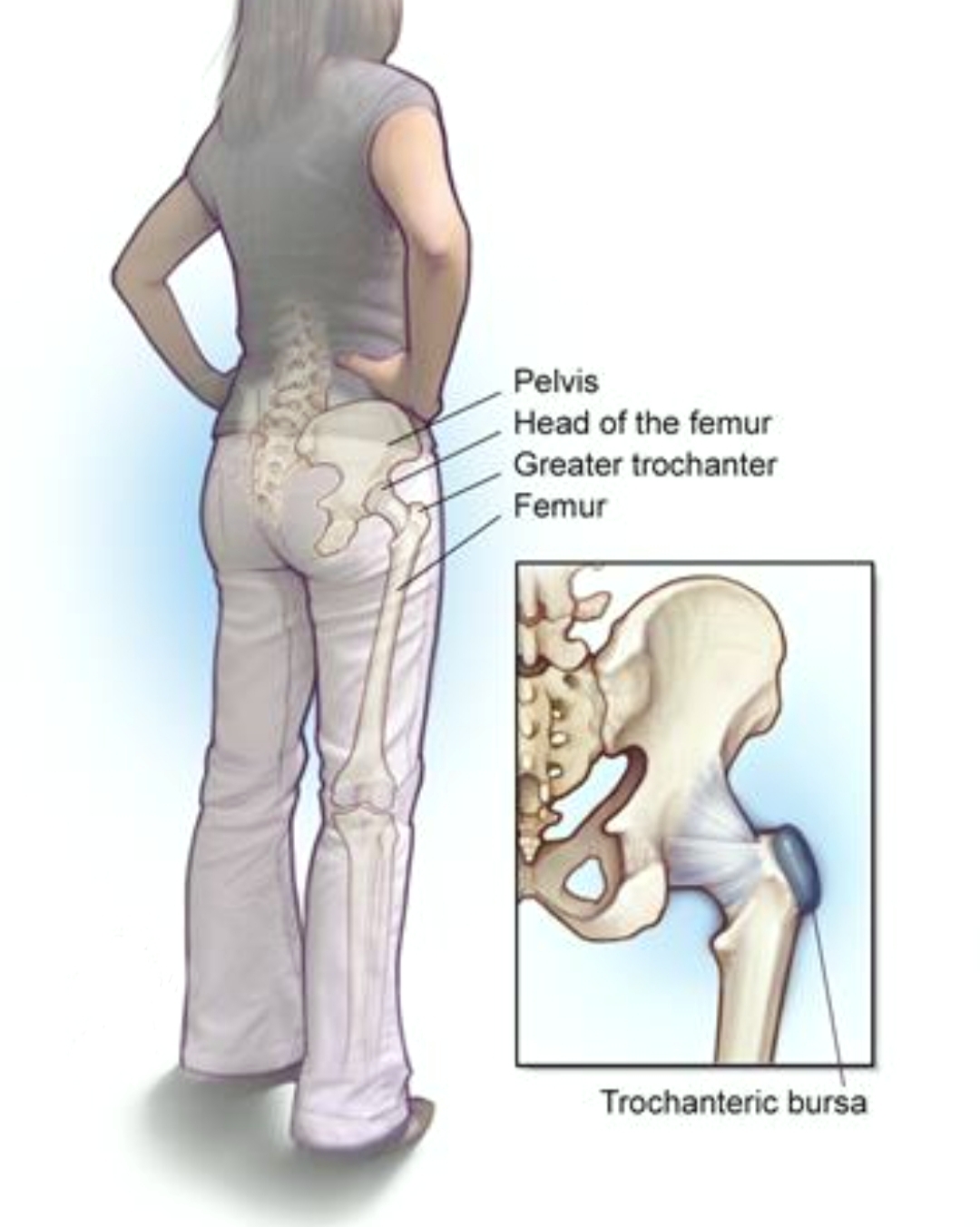
- The Greater Trochanter
- The Upper Margin of the Medial Epicondyle of Femur
- The Line Below the Medial Tibia Condyle
- Tip of Medial Malleolus
- Tip of Lateral Malleolus
These 5 locations would suffice to complete the 4 required measurements in the limb's BPM in this blog.
This is How to use Observation, Olfaction, Inquiry\Auscultation, and Palpation in Paediatrics. (part 10)

The Greater Trochanter
So my next reference point to take would be the upper edge of the Greater Trochanter. The most effective way to search for the Greater Trochanter is to flex the leg to see the Greater Trochanter protrudes to the highest point laterally. Once this point is located, there is one popular acupuncture point situated within one and half-inch diameter from this point called Huan Tiao**, which belongs to the Gall Bladder meridian channel commonly used by TCM practitioner to be listed in this blog. It is a definite good reference point used to locate Huan Tiao Xue. The remaining 5 locations are related to this and therefore the explanations of the femur and tibia bone are necessary.
The next reference point to take note of is the popliteal line or stripe found behind the knee cap posteriorly. This line is formed as a result of flexing of the femur and tibia bone. Please see the diagram. It measures a standard 19 inches from the upper edge or margin of the Greater Trochanter to this popliteal line regardless of size.
The Tip of Medial Malleolus
So, what is Malleolus? It is a bony projection similar to a hammerhead, located on either side of the ankle. Therefore, the identification of these 2 points is very clear now, the tip of the Medial Malleolus is the lower end protruding edge of the tibia bone which is slightly higher than the tip of the Lateral Malleolus.
The Tip of Lateral Malleolus
This tip is the distal end of the fibular bone fused with a fibular notch of the tibia bone to form the lateral ankle bone.
The 2nd required measurement is from the popliteal line to the Tip of the Lateral Malleolus which is standard 16 inches across regardless of size. Please see the diagram.
The Femur
So it is more appropriate to start with the femur bone and its position to identify the position of the Greater Trochanter on the body surface. This is one small part of the Femur bone forming the lateral side of the upper extremity.
In other words, the femur bone has 2 extremities, namely:
- The Upper Extremity
- The Lower Extremity
The Upper Extremity
It contains the femoral head, femur neck and the great trochanter forming the superior epiphysis or the enlarged end part of the shaft. (like plateau)
The femur bone is the proximal bone of the hindlimb in tetrapod vertebrates. The head of the femur articulates with the acetabulum in the pelvic bone forming the hip joint, while the distal part of the femur articulates with the tibia or (shinbone) and patella (kneecap), forming the knee joint.
The Lower Extremity
The lower extremity of the femur or distal extremity is the lower end of the fumer bone in human and other animals, closer to the knee. It is larger than the upper extremity of the femur. It is somewhat cuboid in form but its transverse diameter is greater than its anteroposterior. (Looks like a plateau)
It consists of 2 oblong eminences better known as:
- Lateral epicondyle
- Medial epicondyle
Upper Margin of the Medial Epicondyle
The main function of the Medial Epicondyle is merely for attachment of the ligament to its rough surface. However, our interest here is in TCM perspective is to find the reference point below this epicondyle rough surface when you slide your finger over the skin surface to the point just as the bone taper in to form the angle with a straight femur bone shaft. This is the reference point called the Upper Margin of the Medial Epicondyle of Femur.
The 3rd required measurement is taken from the Upper Edge of the Symphysis Pubis to the Upper Margin of the
Medial Epicondyle is a standard 18 inches across regardless of size.
The Line below the Medial Tibia Condyle
The Tibia bone together with the Fibular bone form the lower limb.
It also has 2 extremities and provides 2 wide platforms or plateau to facilitate weight-bearing force from the body.
At the upper extremity, as the plateau taper at the angle to join the shaft of the Tibia bone, this angle is marked at the Line below the Medial Tibia Condyle.
The 4th required measurement is taken from this reference point to the Tip of the Medial Malleolus which is a standard 13 inches across regardless of size. Please refer to the diagram.
That's it and it wraps up all Bone Proportion Measurement required for the human body, right from the head to the ankle bone. Pat yourself for you had learnt so much now and digested of course.


Best of all
Xiong
***Reference:
( Emperor's Interior Sutra is available on sales better known as the esoteric scripture of the yellow emperor in Amazon, it's just a different way of naming the book cover)
